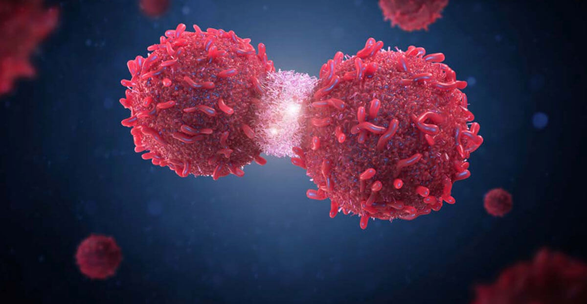
Clinical Research: Phase 1 - Phase 4

Opportunities Beyond Hope: Immuno-Oncology Drug Development

Introduction

Over the past decade, immune-oncology (IO) has become one of the most promising and fastest-growing areas of cancer research and drug development. Present-day advances in immuno-oncology can be attributed to an explosion of research in this area in recent years, leading to a paradigm shift in the understanding of cancer.

Until the late 1990s and early 2000s, cancer was considered a disease of genetic origin, with hallmarks including sustained proliferation, resistance to apoptosis, the ability to promote angiogenesis, and the ability to promote invasion and metastasis. This view failed to consider the dynamic nature of interactions between the tumor and its microenvironment – the normal cells in the surrounding tissue as well as the immune system.

Advances in our understanding of the immune system’s dual role in cancer have led to the development of immunotherapies that target both the tumor and its microenvironment. In this white paper, we explore the immune system’s role in cancer development and the history and challenges of developing immunotherapies for cancer.

Background

The history of cancer immunotherapy dates to the discovery of the dendritic cell in the 1970s. In the early 1990s, Bacillus Calmette-Guérin (BCG), a vaccine used to prevent tuberculosis, was approved as an immunotherapy for early bladder cancer.1 This was followed by the approval of high-dose interleukin (IL)-2 to treat melanoma and renal cell carcinoma (RCC) and the discovery of checkpoint inhibitors.

Recent years have seen an acceleration in the development of IO drugs, with the approval of 22 agents globally. These include nine cancer vaccine products and eight checkpoint modulators. Nineteen of the recently approved IO agents are available in the United States and the European Union, and 13 are available in Japan. The indications with the most marketed IO agents in the U.S. are metastatic melanoma (MM) and non-small cell lung cancer (NSCLC), each of which has six approved products (Figure 1). Notably, hematological malignancies are under-represented among the marketed IO agents.2

Some of the most recently approved IO agents and indications in the U.S. include:3

  • Nivolumab
    • For RCC (in combination with cabozantinib), January 2021
    • For gastric cancer (in combination with chemotherapy), April 2021
    • For esophageal cancer (in combination with chemotherapy and chemoradiotherapy), April and May 2021, respectively
  • Cemiplimab for NSCLC and basal cell carcinoma, February 2021
  • Lisocabtagene maraleucel for large B-cell lymphoma, February 2021
  • Axicabtagene ciloleucel for relapsed or refractory follicular lymphoma, March 2021
    • Idecabtagene vicleucel for relapsed or refractory multiple myeloma, March 2021
  • Dostarlimab for endometrial carcinoma, April 2021
  • Pembrolizumab:
    • For gastric cancer (in combination with trastuzumab and chemotherapy), May 2021
    • For esophageal cancer (in combination with chemotherapy), March 2021

In addition to the large number of approvals in recent years, there are currently more than 4,700 clinical trials of IO agents globally, covering over 500 unique targets in various cancer types. Between 2017 and 2020, the number of IO drugs in development grew by more than 230 percent (Figure 2).4 Of the different types of investigational IO agents, cell therapies constitute the most active area of research with nearly 1,600 ongoing studies of more than 140 pipeline agents, including over 800 clinical trials testing the safety and efficacy of chimeric antigen receptor (CAR) T-cell therapies.2,4

Tumor immunology

A thorough understanding of the underlying tumor biology is critical for the development of IO drugs.

Immune response to tumors

At a high level, tumor immunology can be broken down into three steps (Figure 3):

  1. To initiate immunity, dendritic cells (DCs) must sample antigens derived from the tumor, which can be ingested in situ or delivered exogenously as part of a therapeutic vaccine, making DCs a potential site for therapeutic intervention.

    Upon antigen encounter, the DCs would also have to receive a suitable activation (or maturation) signal, allowing them to differentiate extensively to promote immunity instead of tolerance.
  2. Next, in lymphoid organs, tumor antigen-loaded DCs must generate protective T-cell responses. The precise type of T-cell response needed is unknown but must include the production of CD8+ effector T cells with cytotoxic potential. DCs may also trigger antibody and natural killer/natural killer T (NK/NKT) cell responses, contributing to tumor immunity.

    Tumors go to great lengths to evade the immune response, and systematic studies have identified multiple mechanisms that cancers employ to defeat the immune response:
    • Immunosuppressive cytokines; e.g., transforming growth factor (TGF)-ß, IL-4, IL-6, IL-10
    • Immunosuppressive immune cells; e.g., regulatory T cells (Treg cells) and macrophages
    • Disruption of immune activation signaling; e.g., loss of major histocompatibility complex (MHC) receptor or Indoleamine-dioxygenase production.5,6
  3. Finally, cancer-specific T cells must enter the tumor bed to perform their function. Here, there is the challenge of immune suppression. Presumably by skewing DC maturation, tumors may:
    • Prevent immunization
    • Trigger the “wrong” immune response, or
    • Enable the local accumulation or expansion of Treg cells that would oppose the activity of effector T-cells

Infiltration of Treg cells correlates with poor prognosis in a variety of epithelial tumor types. Tumors may downregulate their expression of MHC Class I molecules or their expression of target tumor antigens. They can also produce a variety of surface molecules, such as programmed death-ligand (PD-L) 1 or PD-L2, which engage receptors on the surfaces of activated T cells (e.g., PD-1 protein), causing T-cell anergy or exhaustion. Expression of such suppressive ligands can be associated with oncogenic mutations seen in many cancers, including phosphatase and tensin homology (PTEN) loss.

Differentiation of CD4+ helper T-cell subsets

Cytokines determine the differentiation of CD4+ helper T-cell subsets.

Many of the IO-targeted therapies activate one of the four main types of T cells:

  • Helper T cells (CD4+)
  • Cytotoxic T cells (CD8+)
  • Suppressor T cells (CD4+CD25+Foxp3+Treg cells)
  • Memory T cells (CD4+ or CD8+CCR7+CD45RO)

CD4+ T cells, which are key regulators of the immune system, differentiate into various T helper (Th) cell lineages with distinct biological functions, depending upon how they are activated.

In the presence of IL-6, IL-21, and TGF-ß, CD4+ T cells differentiate into CD4+ T helper 17 (Th17) cells, a phenotype characterized by expression of the transcription factors retinoic acid receptor-related orphan receptor-gt (RORgt) and signal transducer and activator of transcription 3 (STAT3). IL-1b and IL-23 cytokines can promote and stabilize this phenotype during cell expansion. Once programmed, these Th17 cells secrete IL-17A, IL-17F, IL-21, and IL-22, which play key roles in enhancing autoimmunity and host defense (Figure 4).

Cytokines IL-12, IL-4, and TGF-ß and transcription factors T-bet, GATA3, and FOXP3 have been shown to regulate Th1, Th2, and Treg cell development, respectively. These distinct subsets regulate immune response to foreign, self, and tumor antigens.

Understanding the immunoediting hypothesis

The notion that the immune system not only protects the host against tumor formation but also shapes tumor immunogenicity is the basis of the cancer immunoediting hypothesis.

Accumulated data from animal models and human cancer patients strongly supports the concept that the immune system can identify and control nascent tumor cells in a process called cancer immunosurveillance. In addition, the immune system can promote tumor progression through chronic inflammation, immunoselection of poorly immunogenic variants, and suppression of anti-tumor immunity. Together, the dual host-protective and tumor- promoting actions of immunity are referred to as cancer immunoediting, which is thought to comprise three distinct phases (Figure 5):9,10

  1. Elimination. In this phase, a competent immune system destroys transformed cells.
  2. Equilibrium. Sporadic tumor cells that manage to survive elimination enter this phase, where editing occurs.
  3. Escape. In this phase, immunologically sculpted tumors begin to grow progressively, become clinically apparent, and establish an immunosuppressive tumor.

Development of immunotherapy molecules

The development of immunotherapy molecules over the past 20 years has been an exciting journey, beginning with high-dose IL-2 therapy, one of the first immunotherapies to gain regulatory approval.

High-dose IL-2 therapy

While high-dose IL-2 therapy benefits approximately 6 to 10 percent of patients with MM or RCC, its use is limited by its toxicity and its delivery method.12,13 High-dose IL-2 must be delivered as an inpatient procedure, and use remains limited to select patients treated at experienced centers. Efforts to develop more tolerable regimens have been unsuccessful, and initiatives to better select patients who might benefit from high-dose IL-2 therapy have shown only modest advances. While high-dose IL-2 provides proof of principle that immunotherapy can produce durable benefit in patients with cancer, newer immunotherapies are needed.

Cancer vaccines: current status

Cancer vaccines come in two formats: prophylactic and therapeutic.

Prophylactic (or preventive) vaccines have been used with considerable success in preventing cancers of viral origin, such as hepatitis B virus and human papillomavirus, where the etiologic agent is known. There are currently four approved preventive cancer vaccines for treatment of eight cancer types: anal, cervical, head and neck, vulvar, vaginal, penile, throat, and liver.

In contrast, the development of therapeutic vaccines to treat existing disease has proven problematic.

To date, there are only three approved therapeutic cancer vaccines. The first therapeutic vaccine to be used in practice was BCG, which uses weakened bacteria to stimulate the immune system. BCG is approved for treatment in patients with early-stage bladder cancer. BCG replaced cystectomy as the treatment of choice for carcinoma in situ of the bladder in the mid-1980s. Since that time, other agents have been used in bladder cancer, but none has surpassed the effectiveness of BCG.1

The next approved was sipuleucel-T (marketed as Provenge®), which received FDA approval in April 2010 for treatment of advanced prostate cancer.14 Originally assumed to be an autologous DC-based vaccine, sipuleucel-T comprises an incompletely characterized, complex mixture of peripheral blood mononuclear cells supplemented with a cytokine and tumor-derived differentiation antigen.

The sipuleucel-T Phase 3 clinical results showed little evidence of tumor shrinkage or delay in disease progression. By standard Response Evaluation Criteria in Solid Tumors (RECIST) criteria, only one of the 341 patients in the active arm exhibited a partial response. Nevertheless, investigators observed a 4.1-month improvement in median overall survival (25.8 months vs. 21.7 months), which the FDA deemed significant in a patient population that has few, if any, other effective therapeutic options.15

Most recently, talimogene laherparepvec, or T-VEC (marketed as ImlygicTM), was approved in 2015 for use in recurrent melanoma. T-VEC is an instralesional oncolytic virus therapy made with a form of the herpesvirus. T-VEC selectively targets tumor cells, causing regression in injected lesions, and induces immunologic responses that mediate regression at distant sites.16 Since its initial approval, real-world data have shown response rates of up to 88 percent, with complete response rates as high as 61.5 percent.17

Cancer vaccines: challenges

There are a variety of challenges associated with the development of therapeutic cancer vaccines:

  • The vaccine initially induces an immune reaction against the vaccine itself, not the tumor
  • The antigens are different for each tumor
  • Most immune-responsive tumors auto-vaccinate, but immune regulation prevents an effective response

Based on the aforementioned issues, initial vaccines did not have a major anti-tumor effect in the absence of immune checkpoint control. Data from early vaccine work indicated it was not enough to rely on the virus’s innate ability to kill cancer cells, because the body’s own anti-viral defenses are able to shut them down. Researchers now realize the problem was not so much about oncolysis, but more about what types of transgenes are inserted into the viruses to make the tumor microenvironment stimulatory. Data pulled from Citeline in September 2021 indicate that there are over 50 clinical trials of oncolytic viruses recruiting patients, covering a variety of cancer types.18

Evolution of immune checkpoint inhibitors in solid tumors

The development of immune checkpoint inhibitors is an exciting turning point in IO.

Immune checkpoints refer to a plethora of inhibitory pathways in the immune system that are crucial for maintaining self- tolerance and modulating the duration and amplitude of physiological immune responses in peripheral tissues to minimize collateral tissue damage. It is now clear that tumors co-opt certain immune checkpoint pathways as a major mechanism of immune resistance, particularly against T cells that are specific for tumor antigens (Figure 6).19

T-cell responses are regulated via multiple co-stimulatory and inhibitory interactions. T-cell response to antigen is mediated by peptide-MHC recognized by the T-cell receptor. The B7 family of membrane-bound ligands binds both co-stimulatory and inhibitory receptors. Targeting cytotoxic T-lymphocyte- associated antigen (CTLA)-4 and PD-1 inhibitory receptors has been a major clinical focus.20

Anti-CTLA-4 immunotherapeutic antibodies

Ipilimumab is a member of the novel class of anti-CTLA-4 immunotherapeutic antibodies.

CTLA-4 is a key negative regulator that is recruited to the plasma membrane upon T-cell activation. It binds to members of the B7 family of accessory molecules expressed by DCs and other antigen-presenting cells. CTLA-4 ligation effectively inhibits further activation and expansion, thereby controlling the progress of an immune response and attenuating the chances for chronic autoimmune inflammation. The negative regulation is overcome by use of a blocking antibody, such as ipilimumab.

The fundamental importance of CTLA-4 to controlling T-cell function is well illustrated by the phenotype of CTLA-4 knockout mice, which die of an aggressive lymphoproliferative disorder at a young age. Interestingly, CTLA-4 ligation is also important for the immune-suppressive function of Tregs, further helping dampen T-cell responses. Thus, Treg function is also thought to be blocked by anti-CTLA-4.20

The rationale for using anti-CTLA-4 in cancer therapy was to unrestrain pre-existing anti-cancer T-cell responses and possibly trigger new responses. It is well known that tumor-infiltrating lymphocytes exist for melanoma, and other diseases, and that they can bear specificity for tumor antigens.

In a Phase 3 study, ipilimumab, with or without a glycoprotein 100 (gp100) peptide vaccine, showed improved overall survival compared to gp100 alone in patients with previously treated MM.21 On March 25, 2011, ipilimumab (marketed as Yervoy®) was approved for treatment of unresectable or metastatic melanoma.22

PD-1 pathway inhibitors

Blocking the interaction between the PD-1 protein and one of its ligands, PD-L1, has been reported to generate impressive anti-tumor responses.23

PD-L1 can be expressed on tumor cells either endogenously or induced by adaptive immune resistance.24,25 The interaction between PD-1 and PD-L1 results in T-cell suppression (i.e., anergy, exhaustion, death). In melanoma, renal cell carcinoma, and other tumors, PD-L1 expression has been associated with adverse clinical and pathological features, such as more aggressive disease and shorter survival.26

To date, tumors that have been shown to respond to anti-PD-1 or anti-PD-L1 therapy include breast, cervical, colon, liver, skin, stomach, rectal, melanoma, non-small-cell lung, bladder cancer, and head and neck cancers, as well as RCC, lymphomas, and any solid tumor that is not able to repair errors in DNA. Pembrolizumab (marketed as Keytruda®) and nivolumab (marketed as Opdivo®) were the first of this anti-PD-1 pathway family of checkpoint inhibitors to gain accelerated approval from the FDA for treatment of ipilimumab-refractory melanoma.24 Pembrolizumab has also been approved as a single agent for the first-line treatment of patients with metastatic NSCLC whose tumors have high PD-L1 expression, and nivolumab also has been approved for patients with metastatic squamous NSCLC who have progressed on or after platinum-based chemotherapy.

Rationale for combination therapies

Combination therapies may comprise:

  • Agents that act at the effector stage (e.g., anti-PD-1 or inhibitors of immunosuppression) by re-energizing pre-existing T-cells
  • Agents that act at the proliferation/activation stage (e.g., anti-CTLA-4) to not only enhance pre-existing responses, but also stimulate de novo responses
  • Agents that act on other co-stimulatory or inhibitory pathways
  • Standard of care (e.g., chemotherapy, tyrosine kinase inhibitors, vascular endothelial growth factor inhibitors, radiation therapy)
  • Epigenetic therapy

Combining anti-CTLA-4 with anti-PD1 makes sense biologically, as the two agents remove the brakes from T-cell activation at two distinct stages: proliferation (CTLA-4) and effector function (PD-1). Yet, both might be expected to exhibit similar adverse events, underscoring the need to carefully define the potential for serious toxicity.

In theory, these agents could also work well in conjunction with a vaccine approach, whether exogenous or endogenous.

However, we have yet to see clinical data that support the use of either type of vaccine.

Evidence is emerging that tumor cells can die in multiple ways, with some forms of apoptotic death leading to enhance anti- tumor immune response.

So-called immunogenic cell death is characterized in part by the release of ATP and high-mobility group protein B1, which could activate local-infiltrating myeloid cells and DCs via a purinergic receptor or toll-like receptor 4 (TLR-4), respectively. Cytotoxic agents that elicit this death fingerprint may help induce anti- tumor immune responses and therefore be better candidates for combination therapy with immunologically active agents.

Evaluating response to immunotherapies

It is important to note that, with most immunotherapies, the disease can get worse before it gets better. This is known as pseudoprogression.

Generally, four distinct response patterns are associated with favorable overall survival:27

  1. Response in baseline lesions (i.e., a typical RECIST response)
  2. Stable disease with slow decline in tumor volume
  3. Response following an initial increase in tumor volume
  4. Response following the appearance of new lesions

A clinical challenge with ipilimumab relates to the kinetics of the anti-tumor response. In contrast to conventional cytotoxic therapies that may trigger rapid tumor shrinkage due to direct killing of cancer cells, the stimulation of T-cell responses with ipilimumab may take several months to occur. Tumors may increase in size during this period, and some component of this growth may reflect the consequences of an evolving inflammatory reaction. Indeed, as many as 10 percent of patients treated with ipilimumab who were scored with progressive disease using the modified World Health Organization criteria for tumor size were shown to achieve disease stabilization and prolonged survival.

This unusual pattern of treatment response has led to the proposal of new immune-related response criteria that may aid clinical decision making regarding continuation of therapy.

Introduction of iRECIST

Response and efficacy of oncology agents is measured by RECIST version 1.1 (RECIST 1.1), a set of published rules that define when cancer patients improve, stay the same, or worsen. Unfortunately, these criteria do not easily apply in IO.

As seen with ipilimumab, novel immunotherapeutics trigger different response patterns in tumors than do classic chemotherapy drugs. Applying only RECIST 1.1 to immunotherapy trials can result in:

  • Premature termination of therapy
  • Unnecessary removal of patients from clinical trials
  • Inaccurate interpretation of response

To address questions about the assessment of changes in tumor burden in immunotherapy trials, a consensus guideline – iRECIST – was developed by the RECIST working group, comprising members of industry, academia, the FDA, and the European Medicines Agency. iRECIST calls for the use of modified RECIST 1.1 in cancer immunotherapy trials and describes a standardized approach to solid tumor measurements and definitions for objective change in tumor size for use in such trials.28 iRECIST also introduces a new response criterion called immune unconfirmed progression of disease (iUPD), Figure 8.

When to use iRECIST

The iRECIST criteria allow a standardized response evaluation, considering the relatively rare, but clinically significant, possibility of pseudoprogression within the framework of oncological immunotherapies. For therapy decisions, iRECIST should be used with caution but may offer a good option for systematically documenting therapy outcome. Sponsors should keep in mind that RECIST 1.1 remains the gold standard for defining treatment response-based endpoints in solid tumors for pivotal registration trials. However, iRECIST can be used in conjunction with RECIST 1.1 in later-phase studies and may be used as primary response criteria in exploratory, early-phase studies because assessment is done via RECIST 1.1 until progression.

Conclusion

Immuno-oncology drugs, particularly immune checkpoint inhibitors, can produce durable anti-tumor responses and are changing the landscape of cancer therapy. Successful development of immunotherapies requires a thorough understanding of tumor immunology and careful planning that considers the differences between IO drugs and conventional chemotherapy treatments, including selection of combination therapies, evaluation of disease progression, and management of immune-related adverse events. Despite the obstacles, the future of immunotherapy is a bright one. IO holds the promise of transforming cancer into a more manageable condition with a better prognosis for patients. Researchers are increasingly prioritizing the development of better lab models to study the immune response and the tumor microenvironment. Additionally, technological advances such as liquid biopsies and novel non-invasive imaging strategies also have the potential to improve our ability to discover and validate new biomarkers, making it easier for clinicians to incorporate them into standard clinical practice.29


References

[1] Babalan KN. Bacillus Calmette-Guérin immunotherapy for bladder cancer: overview of BCG immunotherapy. Medscape; 2020. Available at https://emedicine.medscape.com/ article/1950803-overview.

[2] GlobalData, Pharma Intelligence Center. Thematic research: immuno-oncology. GlobalData PLC; 2020.

[3] Cancer Research Institute. FDA approval timeline of active immunotherapies; 2021.

[4] Cancer Research Institute. Immuno-oncology landscape; 2021. Available at https://www.cancerresearch.org/en-us/scientists/immuno-oncology-landscape.

[5] Steinman RM, Cohn ZA. Identification of a novel cell type in peripheral lymphoid organs of mice. I. morphology, quantitation, tissue distribution. J Exp Med. 1973;137(5):1142-1162.

[6] Kirkwood JM, Strawderman MH, Ernstoff MS, Smith TJ, Borden EC, Blum RH. Interferon alfa-2b adjuvant therapy of high-risk resected cutaneous melanoma: the Eastern Cooperative Oncology Group Trial EST 1684. J Clin Oncol. 1996;14(1):7-17.

[7] The Cancer Treatment and Support Foundation. Immunotherapy to cancer; 2021. Available at http://the-cancer-foundation.org/?p=321.

[8] Bailey SR, Nelson MH, Himes RA, Li Z, Mehrotra S, Paulos CM. Th17 cells in cancer: the ultimate identity crisis. Front Immunol. 2014;5:276.

[9] Schreiber RD, Old LJ, Smyth MJ. Cancer immunoediting: integrating immunity’s roles in cancer suppression and promotion. Science. 2011;331(6024):1565-1570.

[10] Mittal D, Gubin MM, Schreiber RD, Smyth MJ. New insights into cancer immunoediting and its three component phases—elimination, equilibrium and escape. Curr Opin Immunol. 2014;27:16-25.

[11] Vesely MD, Kershaw MH, Schreiber RD, Smyth MJ. Natural innate and adaptive immunity to cancer. Annu Rev Immunol. 2011;29:235-271.

[12] Atkins MB, Lotze MT, Dutcher JP, et al. High-dose recombinant interleukin 2 therapy for patients with metastatic melanoma: analysis of 270 patients treated between 1985 and 1993. J Clin Oncol. 1999;17(7):2105-2016.

[13] McDermott DF, Atkins MB. Application of IL-2 and other cytokines in renal cancer. Expert Opin Biol Ther. 2004;4(4):455-468.

[14] National Cancer Institute. Sipuleucel-T; 2019. Available at https://www.cancer.gov/about- cancer/treatment/drugs/sipuleucel-t.

[15] Kantoff PW, Higano CS, Shore ND, et al. Sipuleucel-T immunotherapy for castration- resistant prostate cancer. N Engl J Med. 2010;363(5):411-422.

[16] Harrington KJ, Puzanov I, Hecht JR, et al. Clinical development of talimogene laherparepvec (T-VEC): a modified herpes simplex virus type-1-derived oncolytic immunotherapy. Expert Rev Anticancer Ther. 2015;15(12):1389-403.

[17] Franke V, Berger DMS, Klop WMC, et al. High response rates for T-VEC in early metastatic melanoma (stage IIIB/C-IVM1a). Int J Cancer. 2019;145(4):974-978.

[18] Citeline. Informa Pharma Intelligence; 2021. Available at: https://citeline.informa.com/ trials/results?qId=a24f1464-41bd-4cf0-ab7e-2c1f9869d907.

[19] Pardoll DM. The blockade of immune checkpoints in cancer immunotherapy. Nat Rev Cancer. 2012;12(4):252-264.

[20] Jain N, Nguyen H, Chambers C, Kang J. Dual function of CTLA-4 in regulatory T cells and conventional T cells to prevent multiorgan autoimmunity. Proc Natl Acad Sci USA. 2010;107(4):1524-1528.

[21] Hodi FS, O’Day SJ, McDermott DF, et al. Improved survival with ipilimumab in patients with metastatic melanoma. N Engl J Med. 2010;363 (8):711-723.

[22] National Cancer Institute. Ipilimumab; 2021. Available at https://www.cancer.gov/ about-cancer/treatment/drugs/ipilimumab.

[23] Mahoney KM, Freeman GJ, McDermott DF. The next immune- checkpoint inhibitors: PD-1/PD-L! blockade in melanoma. Clin Ther. 2015;37(4):764-782.

[24] Topalian SL, Drake CG, Pardoll DM. Targeting the PD-1/B7-H1(PD-L1) pathway to activate anti-tumor immunity. Curr Opin Immunol. 2012;24(2):207-212.

[25] Taube JM, Anders RA, Young GD, et al. Colocalization of inflammatory response with B7-h1 expression in in human melanocytic lesions supports an adaptive resistance mechanism of immune escape. Sci Transl Med. 2012;4(127):127ra37.

[26] Thompson RH, Gillett MD, Cheville JC, et al. Costimulatory B7-H1 in renal cell carcinoma patients: indicator of tumor aggressiveness and potential therapeutic target. Proc Natl Acad Sci USA. 2004;101(49):17174-17179.

[27] Wolchok JD, Hoos A, O’Day S, et al. Guidelines for the evaluation of immune therapy activity in solid tumors: immune-related response criteria. Clin Cancer Res. 2009;15(23):7412-7420.

[28] Seymour L, Bogaerts J, Perrone A, et al. iRECIST: guidelines for response criteria for use in trials testing immunotherapeutics. Lancet Oncol. 2017;18(3):e143-152.

[29] Murciano-Goroff YR, Warner AB, Wolchok JD. The future of cancer immunotherapy: microenvironment-targeting combinations. Cell Res. 2020;30(6):507-519.